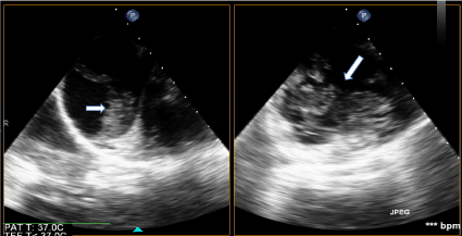


ECMO was complicated by deep vein thrombosis on her left upper femoral superficial vein which resolved on day 13 of admission after anti-coagulation therapy.
#Amniotic fluid embolism second pregnancy serial#
Serial TTEs revealed gradual recovery of heart function and ECMO was successfully weaned off on day 6 of ICU stay with Bakri balloon removed later in the afternoon of the same day. Her coagulopathy resolved over the following 3 days and she was started on IV heparin on day 4 of ICU/ECMO and was converted to fraxiparine on day 7. No heparin administration was started in view of highly deranged coagulopathy. She underwent CT thorax, abdomen and pelvis which reviewed no pulmonary embolus or other etiology to account for postpartum collapse. Prevention of arrhythmia with attention to electrolytes' balance was made.

She was put on anti-heart failure medications. Left superficial femoral artery reperfusion 5F cannula was inserted under ultrasound guidance. 17 F arterial cannula and 21F venous cannula was inserted via left central femoral artery and vein respectively. In view of failure to maintain hemodynamic status with IV adrenaline and dopamine and the appearance of heart failure, the decision was made to support her with extracorporeal membrane oxygenation (ECMO). Intravenous adrenaline at 0.5 mcg/kg/min could only achieve a mean blood pressure of 50 mmHg and a heart rate of 140-150 beats per minute. Emergency transthoracic echo (TTE) on the operating table showed normal right ventricular function and severely impaired left ventricular function with ejection fraction of 10-15% consistent with a stress cardiomyopathy pattern. Her coagulation profile was severely deranged with a PT of 15 seconds and APTT 84 seconds, platelets of 79 × 10 9/l and haemoglobin of 9.5 g/dl. Bakri balloon was inserted to tamponade the uterine-placental surface bleed and to monitor blood loss, with 1.5 litres of blood loss from Bakri drain after 4 hours of resuscitation. IV syntocinon drip (30 units in 1 pint of dextrose saline) was started. Uterotonics such as intravenous (IV) tranexamic acid 1 g, intramuscular (IM) carboprost 250 mcg, and IV duratocin 100 mcg were given. Aggressive management by the multi-disciplinary team including anaesthetist, cardiologist and obstetrician ensured that she was resuscitated with fluids, 6 pints packed cell transfusion, 2 litres fresh frozen plasma, 8 packs of platelet transfusion as well as 6 vials of Recombinant Coagulation Factor VIIa (Novo 7). Patient was brought back to the operating theatre for examination under anaesthesia. She was clinically hypotensive and coagulopathic with bleeding from her nasal and oral cavities and her saturation decelerated to 69%. On examination, there was a persistent ooze of dark red watery blood that seemed poorly oxygenated from her vagina but her uterus was contracted at all times. She developed postpartum haemorrhage 45 minutes post-delivery. She had a second degree perineal tear which was repaired with Vicryl Rapid. Apgar was 9 at 1 minute and 10 at 5 minute. Her second stage of labour lasted 54 minutes and she delivered a baby boy via vacuum assisted delivery for maternal exhaustion. She had spontaneous rupture of membrane at the 13 th hour post prostin and her first stage of labour lasted 6 hours. Oxytocin infusion was started 12 hours later when cervical os was 2 cm and partially effaced. She was admitted at 39 weeks and 3 days for prostin induction for social request. The rest of the antenatal follow-up was uneventful. Non-invasive prenatal screening (Harmony test) at 10 weeks and 5 days was low risk and her screening ultrasound at 22 weeks and 5 days showed a right pelvic kidney. She did not have any significant past medical history of note or drug allergy. We report a 32-year-old Chinese lady, Gravida 1 Para 0, who was booked at 6 weeks and 5 days. In this report we describe the effective value of extracorporeal membrane oxygenation in the successful treatment of a patient with amniotic fluid embolism with uterine conserving management of Bakri balloon treatment of PPH secondary to disseminated intravascular coagulopathy (DIC). We describe our institutional experience with extracorporeal membrane oxygenation (ECMO) use in a pregnant women who presented with amniotic fluid embolism and was complicated by disseminated intravascular coagulopathy and postpartum haemorrhage (PPH) who subsequently developed left ventricular failure requiring ECMO therapy. Amniotic fluid embolism is a rare but catastrophic complication of pregnancy.


 0 kommentar(er)
